Anatomy of the digestive system. Free download Gaivoronsky I. Gaivoronsky I.V., Nichiporuk G.I. Anatomy of the muscular system
skull anatomy
Annotation:
A distinctive feature of this manual is the presence of tables in which all the material presented is synthesized. Many years of teaching experience shows that the material presented in the form of tables is easier to digest and stored in the memory of students in the form of flowcharts necessary for subsequent clinical training. The third edition of the manual has been prepared in accordance with the basic requirements of the curriculum in human anatomy. The latter aspect makes it necessary to work with a textbook and atlases, and the main purpose of this publication is to systematize the information obtained in this way. It provides data on the structure of individual bones of the skull and its integral formations: the eye socket, nasal cavity, cranial fossae, etc.; features of bone development, highlights the differences in the skull of a newborn, characterizes age-related changes, outlines the basics of X-ray anatomy, systematizes data on the contents of integral formations (vessels, nerves, muscles, etc.). When describing the main formations of the skull, along with the Russian names of bone structures, the corresponding Latin terms are given. The illustrations included in this publication make it easier to perceive the material and contribute to its deeper understanding and assimilation. Characterization of the contents of the cavities of the skull and their messages will be useful not only in preparing for the final lesson in craniology, but also for the anatomy exam. The manual is prepared for cadets and students of medical training faculties, students of advanced training faculties, and it can also be used by clinicians of various specialties, in particular, maxillofacial surgeons, neurosurgeons, neuropathologists, ophthalmologists and otorhinolaryngologists.
Anatomy of the digestive system

Annotation:
It meets the basic requirements of the human anatomy curriculum. The manual is designed for cadets and students of medical training faculties, students of advanced training faculties, and it can also be used by clinicians of various specialties. It briefly outlines the fundamentals of the structure of the organs of the digestive system, the features of their blood supply, innervation and lymph outflow. For this purpose, this tutorial has been prepared. When describing the structure of organs, along with Russian names, the corresponding Latin and Greek terms are given. This manual will be useful not only for preparing for the final lesson on the anatomy of the digestive system, but also when repeating the material covered for the exam. The introduction discusses the principles of study and the main patterns of the structure of hollow and parenchymal organs. Due to the fact that the material on the anatomy of the digestive system is studied in the sections "Splanchnology" and "Angioneurology", the integration and systematization of the acquired knowledge is required.
Anatomy of the respiratory system and heart

Annotation:
Along with Russian names, corresponding Latin and Greek terms are given. This manual can be used as a "flow chart" in the study of relevant topics, in preparation for the test in the sections "Splanchnology" and "Angioneurology", as well as when repeating the material covered during the examination session. The publication contains basic information on the anatomy of the respiratory system and heart, briefly outlines the fundamentals of the structure of these organs, the features of their blood supply, innervation and lymph outflow. The manual is designed for students of faculties of training doctors, students of advanced training faculties, and can also be used by doctors of various specialties. The manual has been prepared in accordance with the requirements of the curriculum on human anatomy for higher educational medical institutions.
Anatomy of bone joints

Annotation:
This contributes to easier assimilation of the material and will allow to generalize the knowledge gained in practical classes. The manual is supplemented with elements of kinesiology of the largest and most functionally significant joints. To improve the perception of the material, the manual is illustrated with classic and original drawings. I express my confidence that the presented material on bone joints will be useful to specialists in the field of traumatology and orthopedics, manual therapy, therapeutic physical culture, therapeutic and sports massage. The material is presented briefly, concisely; data on the largest joints are given according to the response scheme traditional for the Department of Normal Anatomy of the Military Medical Academy. The manual has been prepared in accordance with the requirements of the curriculum on human anatomy for medical universities. Data on the innervation and blood supply of bone joints will be useful not only in preparation for practical classes in the 2nd year and for the exam in anatomy, but also for classes in traumatology and orthopedics in 4-5 courses. It contains information on general and particular artrosyndesmology. The manual is designed for cadets and students of medical training faculties, students of advanced training faculties and can be used by clinicians of various specialties.
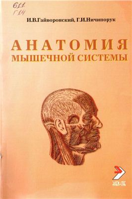
Anatomy of the muscular system

Annotation:
It contains information on general and private myology. This manual presents a description of the main anatomical and topographic formations and their contents. The manual is designed for cadets and students of medical training faculties, students of advanced training faculties and can be used by clinicians of various specialties. The publication contains an original classification of muscles, which provides important principles for clinical anatomy - topographic and the principle of muscle development. As is known, the laying and further formation of muscles is associated with their innervation, therefore, this principle of classification is especially important for the subsequent study of neurology. The manual has been prepared in accordance with the requirements of the curriculum on human anatomy for higher educational medical institutions. The material is presented briefly, concisely, according to the answer scheme adopted at the Department of Normal Anatomy of the Military Medical Academy. These data will subsequently be in demand in the process of studying clinical disciplines, and will also allow generalizing the knowledge gained during training in the first and second years and will contribute to the formation of a holistic view of the structure of the organs of the musculoskeletal system. I express my confidence that the presented material will be useful to specialists in manual therapy and massage.
St. Petersburg: Elbi, 2006. – 64 p.
ISBN: 5-93979-098-4
The textbook meets the basic requirements of the curriculum in human anatomy. It briefly outlines the fundamentals of the structure of the organs of the digestive system, the features of their blood supply, innervation and lymph outflow. When describing the structure of organs, along with Russian names, the corresponding Latin and Greek terms are given. In connection with the introduction of the new International Anatomical Nomenclature (2003), the necessary changes and additions have been made to the manual. This manual will be useful not only for preparing for the final lesson on the anatomy of the digestive system, but also when repeating the material covered for the exam. The manual is designed for cadets and students of medical training faculties, students of advanced training faculties, and it can also be used by clinicians of various specialties.
The ability to download this file is blocked at the request of the copyright holder.
see also

Gaivoronsky I.V., Nichiporuk G.I. Anatomy of the respiratory system and heart
- djvu format
- size 5.86 MB
- added August 28, 2011
Publisher: ELBI-SPb Year: 2010 Pages: 48 The manual was prepared in accordance with the requirements of the curriculum on human anatomy for higher educational medical institutions. The publication contains basic information on the anatomy of the respiratory system and the heart, briefly outlines the fundamentals of the structure of these organs, the features of their blood supply, innervation and lymph outflow. Along with the Russian names, the corresponding Latin and Greek terms are given...
Gaivoronsky I.V., Nichiporuk G.I. Anatomy of the muscular system
- pdf format
- size 9.95 MB
- added December 18, 2011
ELBI-SPb (2005) 84 pages. The manual has been prepared in accordance with the requirements of the curriculum on human anatomy for higher educational medical institutions. It contains information on general and private myology. The material is presented briefly, concisely, according to the answer scheme adopted at the Department of Normal Anatomy of the Military Medical Academy. The publication provides an original classification of muscles, providing important principles for clinical anatomy - top...

- djvu format
- size 8.17 MB
- added February 05, 2012
St. Petersburg: Elbi, 2006. – 64 p. ISBN: 5-93979-098-4 Due to the fact that the material on the anatomy of the digestive system is studied in the sections "Splanchnology" and "Angioneurology", integration and systematization of the acquired knowledge is required. For this purpose, this tutorial has been prepared. It meets the basic requirements of the human anatomy curriculum. It briefly outlines the fundamentals of the structure of the organs of the digestive system, the features of their blood supply ...

Coursework - Anatomical characteristics of the digestive system
Course work- doc format
- size 267.31 KB
- added April 23, 2011
The manual describes in detail and in an accessible language the entire human anatomy. GNI. The value of the nervous system. The general plan of its structure. . Methods for studying the nervous system and VND. Anatomy and physiology of the cardiovascular system, respiratory organs. Anatomy and physiology of the respiratory system. Methods for studying the cardiorespiratory system.
transcript
1 I.V. Gaivoronsky, G.I. Nichiporuk OSTEOLOGY textbook Recommended by the Interuniversity Editorial and Publishing Council for Medical Literature of St. Petersburg as a textbook for faculties of training doctors c. "i St. Petersburg" ELBI-SPb "2005
2 I.v.gayvoroisk, g.i.nichiporuk OSTEOLOGY. Tutorial. SPb.: ELBI-SP S. ISBN Osteology is the first section from which the study of human anatomy begins. It is the most difficult, as it includes a huge number of specific anatomical structures. The latter must be correctly named in Russian and Latin, shown on an anatomical preparation. This manual provides for the systematization of knowledge gained in lectures and practical exercises. For brevity, the main parts of the bones are given under a certain number, and the formations on them are separated by a hyphen and written in the nominative case. Latin terms are given without abbreviations, because in the first semester a very important aspect of learning is the assimilation of anatomical terminology. The text is accompanied by informative illustrations, which show all the necessary anatomical structures. When presenting private issues, special attention is paid to the correct orientation of the bones in relation to the anatomical stance (vertical position of the body, hands turned palms forward). The manual has been prepared in accordance with the requirements of the educational program on human anatomy for higher educational medical institutions. It contains basic information on the anatomy of the skeletal system. I express my confidence that the information presented in this manual will contribute to the successful study of the material in the textbook, will allow you to prepare well for the test and exam, and provide a good basis for self-control and computer testing. ISBN I.V. Gaivoronsky, 2005 G.i. Nichiporuk, 2005 ELBI-SPb, 2005 Signed for printing, Format 60x88 1/16 Offset paper. Headset Times. Volume 4 p. l. Circulation 1000 copies. Order 3373 Publishing house LLC "ELBI-SPB" ID from St. Petersburg, Laboratory pr., 23,
3 GENERAL OSTEOLOGY Osteology is the doctrine of justice. In the body of a young person, there are 206 bones, which, together with their joints, make up the skeleton. Functions of the skeleton 1. Support - the presence of attachment points for soft tissues (muscles, ligaments, fascia, internal organs). its parts in space (bones are levers). 3. Ant 1 1 gravitacs 1 yu iiaya - counteracting the force of gravity. 4. Protective - prevention of damage to vital organs, large vessels and nerves (skull, chest, pelvis). 5. Hematopoietic and immune - the formation of blood cells, the destruction of the pop and in and in the organ and nizm and kr o organizm o in an antibody (provided by red bone marrow). , mainly salts of calcium, phosphorus and other trace elements). and y type p and h n y yu form and structure, characteristic architectonics of vessels and nerves, built 1st, mainly from bone tissue, are covered !! outside the periosteum "1 and containing the bone marrow inside. O steon (Haversian system) is a structural and functional unit of the bone (Fig. 1). It is represented by concentrically arranged bone plates (Haversian), which are in the form of cylinders of different diameters nested into each other , surround the Haversian canal. Between the ostsons are intercalated plates (remaining part of the old octc4) going in all directions. Outside the bones are the general plates. the substance, substantia compacla, is a dense plate covering the bone from the outside, consisting of osteons and bone plates.The diaphysis of tubular bones consists of it (Fig. 2) , and in the form of a thin plate it covers the epiphyses of tubular bones, flat, voluminous and mixed bones.1 2 Fig. 1. Strosins of the bone.I - bone graft; 2 -osteon (reconstruction).
4 Sponge, subxtontia sponglosa, represented by sparsely located bone plates. It is present in the epiphyses of tubular bones and constitutes the main missus of flat and voluminous KOCTcii. The periosteum, periosleum, covers the outside of the bone, except for the places where articular cartilage is located and muscle tendons or ligaments are attached. It plays an important role in the development (growth in thickness) and nutrition of bones. The red Kocriibiit brain, medulla ossetiin nihrci, is located in the cells of the spongy substance; it performs a hematopoietic function, 1 Yellow bone marrow, medulla osseum Jhiva, is present only in an adult; it is located within the marrowy cavity, which is lined from the inside with an endosteum. An untreated bone of an adult contains about 50% water; 16% fat; 12% organic and 22% inorganic substances; the inorganic substance is represented mainly by calcium salts in the form of crystals pschrokh "naiiigi gn, prndayuii" x cosh strength 1 isc. 2. Figure npokchmujn>yogo ei n f and the femur. 1 - epiphysis proximuiis; 2 m eliiphysis; 3 - substantia spongiosa; 4 - substantia compacta; 5 - cavitus medulluris; 6 - diaphysis. and fragility; the organic matter of the bone is represented mainly by the protein ossiiome, which gives the bone flexibility. Classification of bones 1, Pi located in blue: - bones of the skull; - bones of 7 catchers; - bones of the limbs, 2, Three types of skull bones are distinguished according to the stump of the trepiem p roenigo; - diploic (parietal, occipital, lobe bone, lower jaw); - peumatized (temporal, clavicular, ethmoid, frontal bones and upper jaw); - compass (lacrimal, zygomatic, palatine, nasal bones, inferior conch, vomer, hyoid bone), 3. Four types of oblique tulopish and cops are distinguished in shape and sharpness: - tubular bones: a) long (humerus, forearm bones , femur, bones of the head, clavicle); b) short (metacarpal bones, metatarsal bones, finger bones); 4
5 - flat (tpzvpya bone, sternum, scapula, rib); voluminous (carpal bones, tarsal bones); - mixed bones (vertebrae), 4. Pirizshpiyu: primary; develop on the basis of connective tissue (bones of the roof of the skull; zygomatic, palatine, noses; sh, lacrimal bones; upper jaw and couhiiik); - secondary; develop on the basis of cartilage (bones of the trunk and conscience; ethmoid and hyoid bones, lower 1st concha); - mixed (occipital, sphenoid and temporal bones; and jaw). Bone development 1. Primary bones are formed on the basis of connective tissue (conjunctival and bone stages); they ossify according to the endesmal type: by oppositional growth from ossification points from the center to the periphery (bones of the facial skull, bones of the skull roof). 2. Secondary bones develop on the basis of cartilage (connective tissue, cartilaginous and bone stages); - enchondral type of ossification; from the center of the bone to psri (1) sria (bones of the base of the skull, epiphyses of tubular bones, bones of the body); psychoidral type of ossification; the formation of a bone cuff around the cartilaginous anlage (diaphysis of 1 ribbed bones), Mstaepiphyseal cartilage is a layer of cartilage between the JPyphysis and the diaphysis, which is a zone of bone growth in length. - cervical vertebrae, vertebrae cervicales, - 7; - thoracic vertebrae, vertebrae titoracicae, 12; - lumbar vertebrae, vertebrae himbales, - 5; 2) fused vertebrae; - sacrum, os sacrum, - 5; - coccyx, os coccygls, ~ 3-5. General features of free spinal cords Vertebra, vertebra, composed of three main parts (Fig. 3); 1, Body posniikp, corpus vertebrae. 2, Posnoic arc, arctis vertebrae: pedicles of the vertebral arc, peciicidi arcus vertebrae, form the body and the vertebral arch; 5
6 - vertebral foramen, / og / "en vertehrale, limited by the body and arch of the vertebra; openings of all vertebrae form the spinal canal, canalis vertebral is. 3. Processes of the vertebrae, / j / "ocejiiw vertebrae: a) spinous process, processus spinosus, - unpaired; located behind, along the midline; b) transverse process, paired; located in the frontal plane; c) the upper and lower articular processes, proccwiw articularis superior et pmcessus articularis inferior, are paired; The upper vertebral notch, incisura vertebralis superior, is located between the body and the upper articular process. The lower vertebral notch, incisura vertebralis inferior, is located between the body and the lower articular process; it is larger in size than the upper notch. The intervertebral foramen, / ora / pen intervertebrale, is formed by the connection of adjacent vertebrae (vertebral notches); spinal nerves and blood vessels pass through it. Vertebral orifice: - the body of the vertebra is facing forward; - the spinous process is directed backwards; - the upper vertebral notch is directed upwards (slight); - the lower vertebral notch is located below (deep). physical vertebrae; 2 - pcdiculus arcus vertebrae; 3 - procssus articularis superior; 4 - processus articularis inferior; 5 - processus spinosus; 6 - arcus vertebrae; 7 - processus transversus; 8 - foramen vertebrale; 9 - incisura vertebralis inferior; 10 - incisura vertebralis superior.
7 Cervical vertebrae The main distinguishing feature of the cervical vertebrae (vertebrae cervicales) is the presence of a hole in the transverse process, /ora/nep processus transversus; vertebral vessels pass through it. 1) atlas and axial, atlas et axis, (1 and 2 vertebrae) - atypical vertebrae; 2) 3-7 cervical vertebrae are typical vertebrae. Tipchie vertebrae: - vertebral bodies of small size, have a saddle shape; - the vertebral foramen is large, triangular in shape; - groove of the spinal nerve, sulcus nervi spinalis, - runs along the upper surface of the transverse processes (Fig. 4); - anterior and posterior tubercles, tuberculum anterius et posterius, - located in front and behind at the end of the transverse process; - spinous processes are short, directed somewhat downward; forked at the end; - articular processes are short, located obliquely between the frontal and horizontal planes; the upper articular processes are turned back and up, the lower ones forward and down. The anterior tubercle, tuberculum anterius, of the VI vertebra is more developed - the carotid tubercle, tuberculum caroticum, (the common carotid artery is pressed against it during bleeding). VII cervical vertebra - a protruding vertebra, vertebra prominens: its spinous process is longer, thickened at the end; its tip is well felt under the skin. Atypical vertebrae: Atlas, atlas The 1st cervical vertebra lacks a body, spinous and articular processes. It consists of anterior and posterior arches, as well as lateral masses: 1) anterior arch, arcus anterior: - anterior tubercle, tuberculum anterius, located on the outer (front) surface (Fig. 5); - tooth fossa, fo vea dentis, is located on its inner (rear) surface; Rice. 4. Seventh cervical vertebra. 1-tubcrculurn poslcrius; 2 - sulcus nervi spinalis; 3 - tubcrculurn anterius; 4 - proccssus transversus; 5 - corpus vertebrae; 6 - foram en proccssus transversus; 7 - procssus articularis superior; 8 - procssus articularis inferior; 9 - processus spinosus; 10 - foramen vcrtcbrale.
8 2) lateral masses, massae lalemles: - superior articular fossa, / overt articularis superior, oval, deep; serves to connect with the condyles of the occipital bone; - lower articular fossa, fovea articularis inferior, rounded, insignificant in depth; serves for eoediiiii with an axial vertebra; - transverse process, processus iransversus, has / orame / iprocessus transversus ", does not contain grooves of the spinal nerve and tubercles; 3) posterior arch, arcus posterior: posterior tubercle, tuberculum posterius-, - groove of the vertebral artery, sulcus arteriue vertebralis, passes behind the lateral masses on the upper surface of the posterior arch.Axial vertebra, axis odontoid process (tooth), dem, is located on the upper surface of the vertebral body (Fig. b); this is the body of the first cervical vertebra that has moved in the process of development and accreted: a) the apex of the tooth, apex deiitis b) the anterior articular surface iy6a, facies articularis anterior ilentis, connects with the fossa of the tooth of the atlas c) the posterior articular surface of the lip, facies articularis posterior dentis, ~ comes into contact with the transverse ligament of the atlas Fig. 5. Ltlapt 1 processus irnnsvcrsus; 2 - massa Itttoralis; 3 - nrcus anterior; 4 - lubcrculum unterius; 5 - fovea articularis superior: 6 - foram en processus transversus; 7 - sulcus arteriue vertebralis; 8 - arcus posterior; 9 tuberculum posterius; 10 - foramen vertcbrale; 11 - fovea dentis. 8 Ph.D. 6. OCCDOit noidoitok. 1 - processus iransversus; 2 - dens; 3 apex donlis; 4 - facies articularis posterior dentis; 5 - fiicies articularis superior; 6 - foramen proccssus transversus; 7 - processus articularis inferior; 8 - processus spinosus; 9 - foramen vertebrale; 10-arcus vertebrae.
9 - transverse process, processus transversus, has foramen processus transversus; does not contain grooves of the spinal nerve and tubercles; - upper articular surface, /a c /ej articularis superior, - analogue of the superior articular process; serves for articulation with the lower articular surfaces of the lateral masses of the atlas. Thoracic vertebrae The main distinguishing feature of the thoracic vertebrae (vertebrae thoracicae) is the presence of costal fossae and semi-fossae on the vertebral body, as well as costal fossae on the transverse processes: a) a complete costal fossa, fovea coslalis, is located on the body of I, XIXII vertebrae; serves to attach the head of the same name rib; b) the superior costal fossa (half-fossa), / ov ea coslalis superior, is located on the body of the II-X vertebrae (Fig. 7.8); c) the lower costal fossa (half-fossa), fo vea coslalis inferior, located on the body of I-IX vertebrae; d) the upper and lower costal semi-fossae of adjacent vertebrae form a single articular platform for the head of the rib with each other; e) the costal fossa of the transverse process, fovea coslalis processus Iransversus, is located on the transverse process of the 1st vertebrae; - the thoracic vertebrae are larger than the cervical ones; Th, ThiMx СО 1 Г Thv 7. Thoracic vertebra. 1 - proccssus articularis superior; 2 - incisura vcrtcbralis superior; 3 - fovea costaiis superior; 4 - corpus vertebrae; 5 - fovea costaiis inferior; 6 - incisura verlebralis inferior; 7 - processus articularis inferior; 8 - processus spinosus; 9 - processus transversus; 10 - fovea coslalis processus transversus. ThXI-XIl Fig. 8. The layout of the costal fossae. 1 - fovea coslalis; 2 - fovea coslalis inferior; 3 - fovea costaiis superior; 4 - fovea coslalis processus transversus; 5 - processus spinosus; 6 - processus transversus. nine
10 - the height of the bodies of the thoracic vertebrae from the 1st to the CPth gradually increases; their transverse size increases; - articular processes of the thoracic vertebrae stand frontally: the articular surface of the upper vertebrae is turned back, the lower - forward; - transverse processes facing laterally and backwards; - spinous processes of the thoracic vertebrae are longer than those of the cervical; inclined downwards and cranium-like superimposed on each other. Lumbar vertebrae Lumbar vertebrae, vertebrae lumbales, have a massive body (see Fig. 3); on the preparation are determined by the method of exclusion by the absence of foramen processus transversus elfoveae coslales: - the body of the lumbar vertebra has a bean-shaped shape; the height and width of the body gradually increase from the 1st to the V-ro vertebrae; - the articular surfaces of the articular processes are located in the sapptal plane: in the upper processes they are directed medially, in the lower processes - laterally; - transverse processes of the lumbar vertebrae are located in the frontal plane; - spinous processes are short, flat, directed backwards; located almost at the same level with the vertebral body; - vertebral foramen - triangular in shape. The sacrum The sacrum, os sacrum, consists of five fused sacral vertebrae, vertebrae sacrales: 1) the base of the sacrum, basis ossis sacri, - the upper, wide section: - the upper articular process, processus articularis superior, - paired; connects with the lower articular process of the V-ro of the lumbar vertebra; - cape, promontorium, - an anterior protrusion, formed at the junction of the sacrum with the body of the V-ro of the lumbar vertebra; 2) the top of the sacrum, apex ossis sacri: - sacral horn, cornu ^acra / e, - paired; is a rudiment of the lower articular process (Fig. 9); 3) anterior (pelvic) surface, / ac / e5 anterior (pelvina): - transverse lines, Ppeae Iransversae, are formed as a result of fusion of the bodies of the sacral vertebrae; - pelvic sacral openings, foramina sacralia pelvina; 4) back n0bepxh0ctb,yif/c/ei dorsalis-. ten
11 - median sacral crest, crista sacral is mediana, - unpaired; formed by fusion of the spinous processes; - intermediate sacral crest, crista sacralis intermedia, - paired; formed as a result of the fusion of articular processes; - dorsal sacral foramen, vgash/;w sacralia dorsalia; - lateral sacral crest, crista sacralis lateralis, ~ paired; arose during the fusion of the transverse processes; 5) lateral part, pars lateralis: - ear-shaped surface, yas / e5 auricularis, connects to the same name surface of the pelvic bone; - sacral tuberosity, tuberositas sacralis, located posterior to the ear-shaped surface; connects with ligaments to the tuberosity of the pelvic bone. The sacral canal, canalis sacralis, runs throughout the sacrum; in the region of the apex it ends with the sacral fissure, hiatus sacralis. Ornation of the sacrum: - the base of the sacrum is turned upwards; - the tip of the sacrum is directed downward; - forward oriented pelvic surface (concave); - the dorsal surface is turned back (convex, contains sacral crests). A ^ B - pars lateralis; 6 - basis ossis sacri; 7 - processus articularis superior; 8 - facies auricularis; 9 - foramina sacralia dorsalia; 10 - cornu sacrale; 11 - comu coccygis; 12 - hiatus sacralis; 13 - crista sacralis intermedia; 14 - crista sacralis lateralis; 15 - crista sacralis mediana; 16 - tuberositas sacralis. 11
12 Coccyx The coccyx, OS coccygis, consists of 3-5 fused rudimentary vertebrae (Fig. 9): - coccygeal horns, cornua coccygea, are rudiments of the upper articular processes; they connect with ligaments to the sacral horns. Ribs Ribs, coi / ae, depending on the attachment are classified into: 1. True ribs, costae vcrae, - seven pairs of upper ribs (I-VII); cartilaginous parts are connected to the sternum (Fig. 10). 2. False ribs, costae sptm ae,-v \\\-X ribs; attached to the cartilage of the overlying rib, forming the costal arch, arcus costalis. 3. Oscillating ribs, costae Jluctuantes, - XI and XII ribs; end in the muscles of the abdominal wall. The rib consists of cartilage and bone parts; in the latter, the posterior end and body are also distinguished (Fig. 11). CipocHHC ribs: 1. Cartilaginous part (costal cartilage), cartilago costalis, - front, shorter part; 2. The bone part, os costale, is the posterior, longer part: 1) the posterior end, extremitasposterior, includes the head, neck and tubercle: - the rib head, caput costae, connects to the vertebral bodies: - the crest of the rib head, crista capitis costae, - divides the articular surface of the head into two parts (in II - X ribs); ribs I, XI, XII do not have a ridge, because the heads of these ribs articulate with complete fossae on the bodies of the same-named vertebrae; - neck of the rib, sonite costae, - narrow part of the rib; - the tubercle of the rib, tuberculum costae, is located between the neck and the body; - the articular surface of the tubercle of the rib, facies articularis tuberculi costae, serves to connect with the transverse process of the corresponding thoracic vertebra; - protrusion of the tubercle of the rib, eminentia tuberculi costae, - ligaments are attached to it; no tubercles on ribs XI and XII; 2) the body of the rib, corpus costae: - the angle of the rib, angulus costae, corresponds to the bend of the rib; at the first rib it coincides with the tuberculum costae; - groove of the rib, sulcus costae, runs along the lower edge of the rib; it contains blood vessels and nerves; - bodies of P-CHI ribs have inner and outer surfaces; top and bottom edges. 12
13 Oriei of rib gation: - the cartilaginous part of the rib is turned forward; - the head of the rib is directed back; - along the lower edge there is a groove of the rib. Features of the 1st rib: - the body of the 1st rib has an upper and lower surface; medial and lateral edges; - tubercle of the anterior scalene muscle, luberculum musculi scaleiu anlehoris, located on the upper surface of the 1st rib; the muscle of the same name is attached to it; - groove of the subclavian vein, sulcus venae stibclaviae, located in front of the luberculum musculi scaleni anterioris; - groove of the subclavian artery, sulcus arteriae subclaviae, located behind the luberculum musculi scaleni anterioris. Sternum The sternum, sternum, is a flat bone, consisting of three parts - the handle, the body and the xiphoid process (Fig. 10) Fig. 10 10. Chest. 1 - manubrium sterni; 2 - corpus sterni; 3 - processus xiphoideus; 4 - cartilago costalis; 5 - cor) 5us costae; 6 - collum costae; 7 - caput costae; 8 - incisura jugularis; 9 - incisura clavicularis; 10 - costae verac; 11 - costae spuriac; 12 - costae fluctuantes. Rice rib (L): II rib (B). 1 - facies articularis capitis costae; 2 - caput costae; 3 - collum costae; 4 - facies articularis tuberculi costae; 5-angulus costae; 6 - cory^us costae; 7 - sulcus venae subclaviae; 8 - tuberculum m. scaleni anterioris; 9 - sulcus arteriae subclaviac. thirteen
14 1. The handle of the sternum, manubrium sterni: - the jugular notch, incisura Jugularis, is located above, in the middle; - clavicular notch, incisura clavicularis, - steam room; located on the side and top; serves to connect with the collarbone; - rib notch, incisura costal is, - steam room; located on the side; serves to connect with the cartilage of the 1st rib; at the lower edge of the handle is half of the notch for the cartilage of the 2nd rib, incisura costalis II; - the angle of the sternum, angulus sterni, is an anterior protrusion formed when the handle and body are connected. 2. The body of the sternum, corpus sterni: - outer surface, / ac / ej externa, ~ convex; - inner surface, / ac / ej interna, - concave; - rib clippings, incisurae costales, - for cartilage Il-VlI-ro ribs. 3. The xiphoid process, processus xiphoideus, can be bifurcated at the end or has a hole. Orieschity 1 "rudiy: - the handle of the sternum is directed upward; - the xiphoid process is directed downward; - the outer surface is directed forward (convex); - the inner surface is directed backward (concave). 14 SKULL The skeleton of the head is the skull, cranium, includes the following sections: 1) brain skull, cranium cerebrale (neurocranium): - base of the skull, basis cranii; - roof (vault), calvaria; 2) facial skull, cranium viscerale (splanchnocranium). Bones М031Х1Н0Г0 chersia; - occipital bone, os occipitalc, sphenoid bone, os sphenoidale; - frontal bone, os frontale; - ethmoid bone, os ethmoidale\ - parietal bone, os parietale; - temporal bone, os temporale. Bones of the facial skull; - upper jaw, maxilla", - palatine bone, ospalatinum; "zygomatic bone, os zygomaticum \ - nasal bone, os nasale; - lacrimal bone, os lacrimale;
15 - lower nasal concha, concha nasalis inferior; - vomer, vomer, - lower jaw, mandibula "- hyoid bone, os hyoideum. Occipital bone vertebral canal (Fig. 12).It consists of the main and lateral parts, as well as the occipital scales.1. The main part, pa / -5 basilaris: - slope, clivtis, located on the inner surface; 2. Lateral part, pars lateralis, - steam room: - occipital condyle, condylus occipitalis, connects to the upper articular fossa of the atlas; - condylar yasha, fossa condylaris, located behind the condyle; 16 Fig. 12. Occipital bone A - external view B - internal view 1 - condylus occipitalis; 2 - foramen magnum; 3 - fossa condylaris; 4 - crista occipitalis externa; 5 - linea nuchae inferior; 6 - linea nuchae superior; 7 - protuberantia occipitalis extcma; 8 - linea nuchae suprema; 9 - probe in the canalis hypogiossaiis; 10 - tuberculum pharyngeum; 11-clivus; 12 - processus jugularis; 13-pars lateralis; 14-crista occipitalis inlema; 15-protuberanliaoccipitalis interna; 16-sulcussinussagiltalissuperioris; 17-margo lambdoideus; 18 - sulcus sinus transversi; 19 - margo mastoidcus; 20 - sulcus sinus sigmoidci; 21 - tuberculum jugulare. fifteen
16 - condylar canal, canalis condylaris, opens at the bottom of the fossa of the same name; fickle; - canal of the hypoglossal nerve, canalis hypoglossalis, penetrates the condyle in the middle; - jugular notch, incisura Jugularis, located on the outside of the lateral part; - jugular process, processus jugularis, lies behind the jugular notch; - groove of the sigmoid sinus, sulcus sinus sigmoidei, passes medially to the jugular process; - jugular tubercle, tuberculum jugulare, located near the foramen magnum, above the hypoglossal nerve canal. 3. Occipital scales, squama occipitalis: 1) external n0bcpxh0ctb,yac / ei externa "- external occipital protrusion, protuberantia occipitalis externa", - external occipital crest, crista occipitalis externa, descends along the midline down from protuberantia occipitalis externa: upper nuchal line, Npea pisiae superior, - lower nuchal line, Npea nuchae inferior, - the highest nuchal line, Npea nuchae suprema, located above Npea nuchae superior, unstable; - lambdoid edge, margo lambdoideus, located above and laterally; adjacent to the parietal bone; - mastoid margin, margo mastoideus, located below and laterally; connects with the mastoid process of the temporal bone; 2) internal (brain) surface, / I with / ev interna (cerebraus): - cruciform elevation, eminentia crticiformis: a) internal occipital protrusion, protuberantia occipitalis interna, located in the center of eminenlia cruciformis; b) the internal occipital crest, crista occipitalis interna, is located below, continuing to the foramen magnum; c) the groove of the superior sagittal sinus, sulcus sinus sagittalis superioris, goes up from the protuberantia occipitalis interna; d) the groove of the transverse sinus, sulcus sinus transversi, is located on the sides of the protuberantia occipitalis interna. 1 b Orientation of the occipital koci h: - the basilar part is facing forward (the slope is directed upwards); - scales are oriented backwards and upwards; - occipital condyles facing down.
17 Parietal bone Parietal koet, osparietale, - steam room; participates in the formation of the roof of the skull, it distinguishes the outer and inner surfaces; it has four edges and four corners (Fig. 13). 1. Surface of the parietal braid1 and 1) outer surface, / d s / ej externa: - parietal tubercle, tuber parietale\ lower temporal line, Ipea temporalis inferior, runs parallel to the lower (scaly) edge; - the upper temporal line, Ipea temporalis superior, is located above the previous one; 2) inner (brain) surface, / ac / ej interna (^ cerebralis): - groove of the superior sagittal sinus, sulcus sinus sagittalis superioris, located in the region of the upper edge; - the groove of the sigmoid sinus, sulcus sinus sigmoidei, is located in the region of the mastoid angle; - arterial grooves, sulci arteriosi "- groove of the middle meningeal artery, sulcus arteriae meningeae mediae, extends from angulus sphenoidalis upwards and backwards; digital impressions, impressiones digitatae \ - cerebral protrusions, at" 5th cerehralia-. 10 Fig. 13. Parietal bone A - outer surface (left bone); B - inner surface (right bone). 1 - margo squamosus; 2 - angulus sphenoidalis; 3 - linea temporalis inferior; 4 - mnrgo frontalis; 5 - angulus frontalis; 6 - margo sagittalis ; 7 - foramen parietale; 8 - tuber parietale; 9 - angulus occipitalis; 10 - margo occipitalis; 11 - linea temporalis superior: 12 - angulus mastoideus; 13 - sulcus sinus sagittalis superioris; 14 - sulcus sinus sigmoidei; 15 - sulcus arteriae meningeae mediae, 17
18 - dimples vp & wyn ^ mm, foveolaegranulates, located in the region of sulcus sinus sagittalis superioris-, - parietal opening, foram en parietale, located near the sagittal edge; fickle. 2. The edges of the parietal braid: - the sagittal (upper) edge, margo sagittalis (superior), is located above; connects with the same edge of the opposite bone; - frontal (anterior) edge, margo frontalis (anterior), connects to the scales of the frontal bone; - occipital (posterior) edge, margo occipitalis (posterior), connects to the scales of the occipital bone; - scaly (lower) edge, margo squamosus (inferior), connects to the temporal bone. 3. At "ly parietal kocih: - frontal angle, angulusfrontalis, - anterior-superior; - occipital angle, angulus occipitalis, - postero-superior; - wedge-shaped angle, angulus sphenoidalis, - anterior-inferior; - mastoid angle, angulus mastoideus, - postero-inferior Orientation of the parietal braid: - the outer surface (convex) is directed to the lateral side; - the inner surface is directed to the medial side (vogout; arterial grooves pass through it); - the wedge-shaped angle is directed forward and downward (the most acute; on its the inner surface begins a furrow of the middle meningeal artery, which goes from bottom to top and back). Frontal bone Frontal bone, osfrontale, is located in front of the skull. It consists of the frontal scales, orbital and nasal parts: inner (brain) surface, yaciej interna (cerebralis), - concave (Fig. 14): - groove of the superior sagittal sinus, sulcus sinus sagittalis superioris, located in the midline; - forehead the crest, crista frontalis, is located downward from sulcus sinus sagittalis superioris-, - a blind hole, ugate / 1 caecum, is located at the lower end of crista frontalis "- arterial grooves, sutci arteriosi; - finger impressions, impressiones digitatae", 1 8
19 - cerebral BbicTynbi, 7 "ga cerebralia \\ - dimples grgl \ ulyapsh, / o\ eo1ae gramilares, located in the sukus sinus sagittaiis superions \ 2) outer surface, yi / ciei externa, - convex: - supraorbital margin, margo supraorbitalis, separates the scales from the orbital part; - supraorbital notch (supraorbital foramen), incisura supraorbitalis (foramen supraorbitale) is located at the medial end of margo supraorbitalis; - zygomatic process, processus zygomaticus, is a continuation of margo supraorbitalis in the lateral direction; - the temporal line, Ipea temporalis, runs up and back from the processus zygomaticus; - superciliary arch, arcus superciliaris, located above the medial part of margo supraorbitalis; - frontal tubercle, tuberfrontale-, - nose bridge, glabella, located between the superciliary arches. 2. The orbital part, p arsogy1ash, - part I: 1) the cerebral surface, uyas / e5 cerebral is \ - finger impressions, impressiones digitatae; - cerebral protrusions, 7mga cerebralia \\ In Gb 15 "9 Fig. 14. Frontal bone. A - outer surface; B - inner surface. 1- sulcus sinus sagittaiis superioris; 2 - crista frontalis; 3 - linea temporalis; 4 - fossa 5 - sinus frontalis; 6 - incisura ethmoidalis; 7 - fovea irochlcaris; 8 - foramen supraorbitale; 9 - pars orbitalis; 10 - processus zygomaticus; 11 - foveolae cthmoidales; 12 - squam a frontalis; 13 - tuber frontale; glabella : 15-spina nasalis; 16-pars nasalis; arcus supcrciliaris; 18 - margo supraorbitalis. 19
20 2) orbital (lower) surface, / I with / ei orbitalis (inferior) -. - the fossa of the lacrimal gland, fossa glandulae lacrimalis, is located laterally, in the region of the zygomatic process; - block fossa, fovea trochlearls, located on the medial side; - block spine, spina trochlearls, - bones! the spine within the fovea irochlearis-, is the place of fixation of the tendon of the superior oblique muscle of the eyeball; - lattice dimples, / oveo / ae ethmoidales, are located at the medial edge of the orbital part; cover the cells of the labyrinth of the ethmoid bone; - anterior and posterior ethmoid openings (notches), foramina ethmoidalla aiuerlus et posterius (Incisurae ethmoidales anterior et posterior), located at the medial edge of the orbital part, on the medial wall of the orbit. 3. Nasal part, / jarj nasalis: - lattice notch, incisura ethmoidalis, located behind the pars nasalis, between the orbital parts; - nasal spine, spina nasalis, - a protrusion downward, located in the middle of the nasal part; - the frontal sinus, sinus frontalis, is divided by a septum (septum sinuum frontalium) into two asymmetrical parts; can be multi-chamber; sometimes absent; - openings of the frontal sinus, aperturae sinuum frontalium, located near the spina nasalis. Orientations of the frontal bone: - the outer surface is directed forward (frontal tubercles, superciliary arches); - backward facing the inner surface (groove of the superior sagittal sinus); - the nasal spine is directed downwards. Ethmoid bone The ethmoid bone, os ethmoidale, fills the eponymous notch of the frontal bone. It consists of perforated and perpendicular plates, as well as a lattice labyrinth (Fig. 15). A perforated (trellised) plate, lamina cribrosa, separates the nasal cavity and the anterior cranial fossa. On it are located: - cockscomb, crista galli", - wings of a cockscomb, alae crlstae galli, are in front; - a blind hole, foramen caecum, is limited by alae cristae galli, (sometimes it lies entirely in the frontal bone); 20
21 - openings of the cribriform plate, cribrae laminae cnbrosae\ olfactory nerves penetrate through them from the nasal cavity, pp. olfactorii. 2. Lattice libyrinth, labynnthus ethmoidalis, - paired; - orbital plate, lamina orbitalis, - solid; located on the lateral side; is part of the medial wall of the orbit; - cells of the ethmoid bone, cellulae ethmoidales, (anterior, middle and posterior, cellulae etmoiclales anteriores, medii etposteriores) - these are cavities of the ethmoid labyrinth; - ethmoid vesicle, bulla ethmoidalis, - the largest cell of the ethmoid bone; localized in the posterior part of the labyrinth; - superior nasal concha, concha nasalis superior, - small; located on the medial surface of the labyrinth in its posterior part; - middle nasal concha, concha nasalis media, larger than the previous one; located under it; - the highest nasal concha, concha nasalis suprema, is located above the concha nasalis superior, unstable; - the upper nasal passage, meatus nasi medius, is located between the upper and middle nasal conchas; - middle nasal passage, meatus nasi medius, located under the middle nasal concha; - hook-shaped process, processus uncinatus, - an outgrowth at the posterior end of the middle turbinate; fickle; - ethmoidal funnel, infundibulum etlmoidale, opens between the processus uncinatus et bulla ethmoidalis-, connects the frontal sinus and the middle nasal passage. 3. Perpendicular plate, lamina perpendicularis, is part of the bony septum of the nose; connects with the nasal bones, the vomer and the sphenoid bone (crista sphenoidalis), as well as with the cartilaginous septum of the nose. Orientation of the solute braid; - a cockscomb is directed up and forward; - the perforated plate is directed upwards; - perpendicular plate facing down. Rice. 15. Ethmoid bone. 1 - crista galli; 2 - ala crista galli; 3 - lamina cribrosa; 4 - cellulae ethmoidales; 5 - labyriilhus ethmoidalis; 6 - lamina penendicularis; 7 - concha nasalis media; 8 - concha nasalis superior; 9 - concha nasalis suprema; 10 - lamina orbitalis. 21
22 Sphenoid bone The sphenoid bone, os sphenoidale, lies in the center of the base of the skull. It consists of a body, paired - small and large wings, as well as a paired pterygoid process (Fig. 16.17). 1. The body of the wedge-shaped kosgi, corpus ossis sphenoidalis, is located in the middle; located on the body: - Turkish saddle, sella turcica: a) pituitary fossa, / ossa hypophysialis, - this is a depression in the center of the Turkish saddle, which is a receptacle for the pituitary gland; Rice. 16. Sphenoid bone. A - top view; B - rear view. 1 - a1a minor; 2 - sulcus prechiasmatis; 3 - tuberculum sellae; 4 - fossa hypophysialis; 5 - canalis opticus; 6 - fissura orbitalis superior; 7 - foramen rotundum; 8 - facies cerebralis; 9 - foramen spinosum; 10 - foramen ovale; 11 - sulcus caroticus; 12 - dorsum sellae; 13 - tuberculum clinoideum posterius; 14 - lingula sphenoidalis; 15 - tuberculum citnotdeum anterius; 16-alam ajor; 1 7 -fossa scs^hoidea; 18-lamina lateralis; 19-lamina medialis; 20 - rostrum sphenoidale; 21 - sophysis ossis sphenoidalis; 22 - procssus plerygoidcus; 23 - hamulus pterygoideus; 24 - fossa pterygoidea; 25 - canalis pterygoideus. 22
23 b) the back of the Turkish saddle, dorsum sellae, limits the pituitary fossa from behind; has a paired outgrowth facing up and forward - the posterior inclined process, tuberculum dinoideum posterius \ c) tubercle of the saddle, tuberculum sellae, limits the pituitary fossa in front; - the precrossing sulcus, sulcus prechiasmatis, is located in front of the tuberculum sellae", it contains the optic chiasm; - the carotid sulcus, sulcus caroticus, is located on the lateral side of the sella turcica-, the internal carotid artery passes through it; - sphenoid uvula, lingula sphenoidalis, limits the sidcus caroticus laterally and posteriorly; - the sphenoid ridge, crista sphenoidalis, is located on the anterior surface of the body of the sphenoid bone; - the sphenoid shells, conchae sphenoidales, are located on the sides of the crista sphenoidalis", - the aperture of the sphenoid sinus, apertura sinus sphenoidalis, opens on the anterior body surface; - the sphenoid sinus, sinus sphenoidalis, is divided into two asymmetrical halves by a septum (septum sinuum sphenoidalium); can be multi-chamber; - wedge-shaped keel, rostrum sphenoidale, is a continuation of crista sphenoidalis on the lower surface. 2. Small wing, ala minor, - paired: - visual canal, sapash optiats, located at the base of the small wing; - superior orbital vannb, fissura orbitalis superior, located between small and large wings; - anterior inclined process, tuberculum dinoideum anterius, paired; turned back, located laterally in relation to the sidcus caroticus. 3. Big wing, ala major. On top of a “large wing 1) the cerebral surface, / ac / ej cerebralis, is concave; facing the cranial cavity: - finger impressions, impressiones digitatae; - cerebral protrusions, yi / ga cerebraliw, - a round hole, / ogatei rotundum; - oval hole, vora / un ovale; - spinous opening, foramen spinosum ", 2) orbital surface, yas / e5 orbitalis, facing the orbit; 23
24 3) temporal surface, / a s / ej temporalis: - infratemporal crest, crista infratemporalis, located in its lower part; 4) the maxillary surface, /ac/e^ maxillaris, is directed towards the tubercle of the upper jaw. The edges of the large wing - the scaly (posterior) edge, margo squamosus (posterior), connects to the scales of the temporal bone; - zygomatic (anterior) edge, margo zygomaticus (anterior), separates the temporal surface from the orbital; connects with the zygomatic bone; - frontal edge, margo frontalis, located above and in front; connects to the bone of the same name; - the parietal edge, margo parietalis, connects to the bone of the same name. 4. Wings; 1st process, rgastklpterygoidexis, - paired; the body departs vertically downward at the base of the large wings: - the pterygoid canal, canalispterygoideus, pierces it at the base; - medial plate, lamina medialis", - lateral plate, lamina lateralis-, - navicular t cl, fossa scaphoidea, - a slight expansion in the upper part of the medial plate; Fig. 17. Sphenoid bone. Front view. 1 - margo parietalis; 2 - ala major; 3 - ala minor; 4 - canalis opticus; 5 - concha sphenoidalis; 6 - sinus sphenoidalis; 7 - fissura orbitalis superior; 8 - facies orbitalis; 9 - margo frontalis; 10 - margo squamosus; 11 - margo zygomaticus; 12 - facies maxillaris; 13 - incisura pterygoidca; 14 - lamina medialis; 15 - canalis pterygoideus; 16 - crista sphenoidalis; 17 - hamulus pterygoideus; 18 - lamina lateralis; 19 - foramen rotundum; 20 - crista infratemporalis; 21 - facies temporalis. 24
25 - pterygoid hook, hamuhispte) ygoicleiis, - outgrowth in the lower part of the medial plate; - pterygoid fossa, fossa pterygoidca, located behind, between the lateral and medial plates; - wing notch, incisura pterygoldea, located below between the plates; it includes the pyramidal process of the palatine bone; - a large palatine sulcus, sulcus palatiints major, runs along the anterior edge of the pterygoid process. Orientation of the sphenoid bone: - the pterygoid processes are directed downward; - the pituitary fossa is turned upwards; - on the front surface of the body, the aperture of the sphenoid sinus opens; the back of the Turkish saddle is turned back; Temporal bone Temporal bone, os temporale, - steam room; located between the occipital and sphenoid bones; contains the organ of hearing and balance; the most important vessels and nerves pass through it. The temporal bone consists of petrous, tympanic, mastoid and squamous parts (Fig. 18,19). 1. The stony part (shchmits.a), par5pelivsa (pyramis), has the shape of a three-sided pyramid; it has an apex and a base; front, back and bottom surfaces; anterior, posterior, and upper edges, Chasgi 11iram1i s 1) the apex of the pyramid, apex pyramis, faces forward and medially; in the area of \u200b\u200bthe apex open: - the internal carotid opening, / drame / i sagochsit internum, - the musculo-tubal canal, canalis musculotubarius ", 2) the base of the pyramid, basis pyramis, is covered by the tympanic part. The surfaces of the pyramid and the formations located on them 1) the front surface facies anterior, facing the middle cranial fossa: - stony-scaly fissure, // l^ngya petrosquamosa, separates the pyramid from the scales; - trigeminal depression, impressio trigemini, located closer to the top of the pyramid (the trigeminal ganglion is adjacent); - cleft canal large stony nerve, hiatus canalis n. pelrosi majoris, is located medially; continues into the sulcus of the same name (sulcus n. petrosi majoris) ". 25
26 - cleft canal of the small stony nerve, hiatus canalis n. petrosi minoris, located laterally; continues into the furrow of the same name (sulcus n. pelrosi minoris); - arcuate rise, eminentia arcuata, located in the middle of the front surface; corresponds to the anterior semicircular canal of the labyrinth; - the roof of the tympanic cavity, legmen lympani, is a platform lying behind the eminentia arcuala; 2) the posterior surface, yas / e5posterior, faces the posterior cranial fossa: - the internal auditory opening, porus acusticus internus, leads to the internal auditory canal, meatus acusticus internus (facial and precillary nerves pass); - subarc fossa, fossa subarcuata, located posterior to porus acusticus internus, under eminentia arcuata; - external opening of the vestibule aqueduct, apertura externa aqueclucius veslibuli, - a slight gap; lying lateral and below the fossa subarcuata; 3) the lower surface of the pyramid id, y a c / e i inferior, faces the outer base of the skull: 12 Fig. 18. Temporal bone (right). A - outside view; B - inside view. 1 - squama temporalis; 2 - processus zygomaticus; 3 - fossa mandibularis; 4 - fissura tympanosquamosa; 5 - procssus styloidcus; 6 - pars tyrapanica; 7 - porus acusticus externus; 8 - pars mastoidea; 9 - incisura mastoidea; 10 - foramen mastoideum; 11 - incisura mastoidea; 12 - porus acusticus intcmus; 13 - eminentia arcuata; 14 - legmen tympani; 15 - sulcus sinus petrosi superioris; 16 - sulcus sinus sigmoidei; 17 - apertura externa aqueductus vestibuli; 18 - fossa subarcuata; 19 - sulcus sinus petrosi inferioris; 20 - apex pyramis; 21 - impressio trigemini. 26
27 - jugular sc &, fossajugiilaris, located in the back of the facies inferior; - external carotid hole, foram en carolicum externum, located in front of the fossa Jugularis \ leads to the carotid canal, canalis caroticus, in which the internal carotid artery passes; - a stony dimple, /b and "/ya pelmsa, is a small depression located in the scallop separating foramen carolic um exlemum from fossa jugiilaris; - the lower opening of the tympanic tubule, apetiura inferior canaliculi tympanici, opens at the bottom of the fossula petrosa; - styloid process, processus styloideus-, - styloid foramen, stylomasloideum styloid, located between processus slyloideus and processus masloideus. The edges of the pyramid and the formations located on them 1) the front edge, maigo anterior, is located on the lateral side of the top of the pyramid; under it, the muscular-tubal canal opens; 2) the upper edge, margo superior, separates the anterior and posterior surfaces; - furrow of the superior stone sinus, sulcus sinus petrosis superioris, runs along the margo superior, 3) posterior margin, margo posterior, separates the posterior and inferior surfaces; - groove of the lower stony sinus, sulcus sinus petrosi inferioris, runs along the margo posterior; Rice. 19. Temporal bone (right). Bottom view. I - fissura pctrosqiiam osa; 2 - fissura pclrotympanica; 3 - canalis musciilotubarius; 4 - foramen caroticum inlernum; 5 - apex pyramis; 6 - foramen caroticum externum; 7 - sulcus sinus petrosi inferioris; 8 - fossula petrosa; 9 - apcrtura externa canaliculi cochleae; foramen canaliculi mastoidei; II - fossa jugularis; 12 - foramen stylom astoideum; 13 - sulcus arteriae occipitalis; 14 - incisura mastoidea; 15 ~ processus mastoidcus; 16 - porus acusticus exiernus; 17-pars tympanica; 18-processus styloidcus; 19 - fossa mandibularis; 20 - tuberculum articulare; 21 - processus zygomaticus. 27
28 - external opening of the cochlear tubule, arroma externa canaliciili cochleae, - a small depression in the region of the posterior end of the margo posterior. 2. Vyribaptya part, pars tympaiuca, limits the external auditory canal from the outside; - external auditory opening, porus acusticus externus, leads to the external auditory canal, meatus acusticus externus, which opens into the tympanic cavity, cavitas tympanica-, - tympanic mastoid manb, fissura tympanomastoidea, separates /; I / ^ tympanica and pars mastoidea; tympanic-squamous uienb, /lssura tympanosquamosa, separates rag ^ tympanica and pars squamosa; part of the pars petrosa is wedged into it, so it is divided into: b) rocky-drum mcnb, / lssurapetrotympanica (a drum string, chorda tympani, emerges from it). 3. The mastoid part, mastoidea, is located behind the external auditory canal: - mastoid process, processus mastoideus; - parietal notch, incisura parietalis, located between the scales and the mastoid process; the mastoid angle of the parietal bone enters it; - mastoid notch, incisura mastoidea, limits the mastoid process from the medial side; - groove of the occipital artery, sulcus arteriae occipitalis, located between the incisura mastoidea and the medial edge of the mastoid part; - mastoid opening, / bgyatel mastoideum, located on the posterior surface of the mastoid process; fickle; - the groove of the sigmoid sinus, sulcus sinus sigmoidei, is located on the medial surface of the pars mastoidea; the mastoideum will open into it; - mastoid cells, cellulae mastoideae, - cavities of the mastoid process; - mastoid cave, antrum mastoideum, - the largest cell; communicates with the tympanic cavity. 4. The scaly part, pars squamosa, is part of the roof of the skull: 1) the brain (inner) surface, / ac / e5 cerebralis (interna) \ arterial grooves, sulci arteriosi "- finger impressions, impressiones digitatae \ - cerebral in s temporal (outer) surface, facies temporalis (externa): zygomatic process, processus zygomaticus", mandibular fossa, fossa mandibularis, takes part in the formation of the temporomandibular joint; 28
29 - articular tubercle, tuberculum articulare, located in front, / own mandibularis. Orientation of the temporal osgn: - forward and medially facing the top of the pyramid; - the mastoid process is directed back and down; - the external auditory meatus is turned to the lateral side; - the internal auditory canal is directed to the medial side; - an arcuate elevation is turned upwards; - the styloid process is directed downwards. Canals of the temporal bone The facial canal, canalis facialis, begins at the bottom of the mealus acusticus iiuenms, then it goes laterally in the thickness of the pyramid (Fig. 20) to the hiatus canalis iien ipelrosi majoris, where it forms a bend at an angle of 90 degrees - the knee of the canalis facialis, genicuhtm canalis facialis . Further, the canal in the horizontal plane goes back and, bending around the tympanic cavity, turns vertically down, ending with the foramen stylomastoideum. From the facial canal, the canal of the large stony nerve and the tubule of the tympanic string branch off. The canal of the large stony nerve, canalis n. pelrosi majoris, starts from canalis facialis in the region of ge / k "sk / and / and canalis facialis, opening on the anterior surface of the pyramid of the temporal bone through hiatus canalis n. petrosi majoris. Drum string tubule, canaliculus chordae tympani, starts from canalis facialis several foramen stylomastoideum, passes through the tympanic cavity and ends within the stony-tympanic fissure, fissura petrotympanica.The tympanic tubule, canaliculus tympanicus, begins b / ossiilapetrosa through apertura inferior canalicuu tympanici; passes through the tympanic Fig. 20. Kyaialy temporal bone I - antrum mastoidcum; 2 - tegmcn tympnni; 3 - canalis semicircularis lateralis; 4 - canalis semicircularis anterior; 5 - hiatus canalis nervi petrosi majoris; 6 - hiatus canalis nervi petrosi minoris; 7 - impressio trigemini; 8 - foramen caroticum internum; 9 - foramen cnroticum extcmum; Yu -eond in canalis facialis; II - foramen stylomastoideum ; 12 - cellulac mastoideae. 29
Rice. 1. Skull: a brain region of the skull (solid line shows the border between the vault and base); b facial part of the skull Lesson 2 HEAD SKELETON. BRAIN AND FACIAL SKULL. SKULL BONES:
Frontal bone Frontal scales squama frontalis Outer surface of facies externa Inner surface of facies interna Supraorbital margin margo supraorbitalis Supraorbital foramen foramen supraorbitalis
Lesson 3. BONES OF THE FACIAL DEPARTMENT OF THE SKULL: UPPER AND LOWER JAWS, PALATINE BONE, VOMOTHER, JOYOGANY BONE, LOWER NOSE CONCH, NASAL BONE, Lacrimal Bone, Hyoid Bone. SKULL IN GENERAL: BRAIN AND
General information about the structure of the human skull. The skeleton of the head is made up of paired and unpaired bones, which together are called the skull, cranium. Some of the bones of the skull are spongy, others are mixed.
1 Boyanovich Yu. V. Atlas of human anatomy / Yu. V. Boyanovich, N. P. Balakirev. Rostov n / a: Phoenix, Kharkov: Torsing, 2005. 734, p. (Medicine for you). PRIVATE OSTEOLOGY Trunk Skeleton Trunk Skeleton
Questions of the theoretical part FINAL LESSON on CRANIOLOGY 1. The development of the skull in ontogenesis. Features of the development of the bones of the brain and facial parts of the skull. Variants of the norm and the main anomalies of development
UDC 611(075.32) BBK 28.706ya723 L34 Original layout prepared by the M-Press studio Design by the Tri Kota design studio L34 Lyovkin, S.S. Atlas of human anatomy / S.S. Levkin. Moscow: AST: 2015. 512
HIGHER PROFESSIONAL EDUCATION M. R. SAPIN, D. B. NIKITYUK HEAD AND NECK ANATOMY Recommended by the State Educational Institution of Higher Professional Education "Moscow Medical
CONTENTS SKULL... Development of the skull... Development of the brain skull... Development of the facial skull... Bones of the brain skull... Occipital bone... Parietal bone... Frontal bone... Ethmoid bone... 0 Temporal
Test tasks in the discipline "Human Anatomy and Physiology" for students of the specialties "Nursing", "Obstetrics" on the topic: "Functional anatomy of bones and their joints" #As part of the skeleton
Skeleton 8 cells FUNCTIONS OF THE SKELETON AND MUSCLES LOCOMOTOR SYSTEM (BONE AND MUSCULAR) ACTIVE PART (Muscles) PASSIVE PART (Skeleton) Energetic Propulsion Protective Hematopoietic Exchange Support Formative
DISCIPLINE "LATIN" SEMESTER I MODULE 1. Introduction to anatomical and histological terminology. The list of competency elements to be formed: Code
MINISTRY OF HEALTH OF RUSSIA State budgetary educational institution of higher professional education IRKUTSK STATE MEDICAL UNIVERSITY (GBOU VPO IGMU of the Ministry of Health of Russia) General Medicine Faculty
MINISTRY OF HEALTH OF THE REPUBLIC OF BELARUS INSTITUTION OF EDUCATION "GOMEL STATE MEDICAL UNIVERSITY" Department of human anatomy with a course of operative surgery and topographic anatomy
PRACTICAL LESSON 1 TOPIC: Anatomy as a science. Axes and planes. Anatomical terminology. General osteosyndesmology. Bones of the spinal column and chest and their connection. Spine as a whole.
Laboratory work STRUCTURE OF THE AXIAL SKELETON Purpose of the work: to study the structure of the bones of the human axial skeleton: the structure of the vertebrae, bones of the chest and bones of the skull, to consider the structure on dummies, posters
MINISTRY OF HEALTH OF UKRAINE KHARKIV NATIONAL MEDICAL UNIVERSITY Department of Human Anatomy Head of Department Professor A. A. Tereshchenko MATERIALS ON HUMAN ANATOMY for preparation
SECTION Introduction Duration of the study of the section 3 hours of practical training The purpose of studying the content of the section is an introduction to anatomy, its subject and content, determining its place in a number of biomedical
D.V. Bazhenov V.M. Kalinichenko HEAD AND NECK ANATOMY INTRODUCTION TO CLINICAL ANATOMY TEACHING AID Ministry of Education and Science of the Russian Federation Recommended by the First Moscow State Medical
MINISTRY OF EDUCATION AND SCIENCE OF THE RUSSIAN FEDERATION NOVOSIBIRSK STATE UNIVERSITY Faculty of Natural Sciences Department of Physiology L. A. Obukhova, N. N. Chevagina HUMAN ANATOMY FUNCTIONAL ANATOMY OF THE SKULL
SECTION Introduction to anatomy Duration of the study of the section 2.5 hours of practical training The purpose of studying the content of the section is to study general anatomical terms, areas and parts of the body, general terms
V.T. Palchun DISEASES OF THE EAR, THROAT AND NOSE A TEXTBOOK FOR MEDICAL SCHOOLS AND COLLEGES 2nd edition, revised and supplemented by the Ministry of Education and Science of the Russian Federation
Topic: Skeleton Tasks: To study the parts of the skeleton and the bones that form them Structure of the skeleton The skeleton consists of interconnected bones. It provides our body with support and shape retention, and also protects
220 Surgical anatomy of the head and neck Temporal fossa Superior boundaries: along the superior temporal line. lower: zygomatic arch (symbol) its bottom consists of the bones of the lateral surface of the skull, including
Ministry of Education of the Republic of Belarus Educational Institution "International State Ecological University named after A. D. Sakharov" Faculty of Environmental Medicine Department of Human Biology
PRACTICAL EXERCISES on the musculoskeletal system BONES OF THE TRUNK Be able to show the body, the arch of the vertebrae, the upper and lower vertebral notches, the vertebral foramen and the vertebral
Atop CHM to PDF Converter 2.1 http://www.chmconverter.com NOTE: Full version does not display this page! Anatomy of the skeleton. Human anatomy in diagrams and drawings. Atlas-allowance. N. V. Krylova, I. A. Iskrenko.
OSTEOLOGY Teaching about bones Musculoskeletal system Changing the position of body parts and moving it in space occurs with the participation of: (1) bones that act as levers; (2) skeletal muscles,
Rice. Bone development: a - cartilaginous stage; b - the beginning of ossification: 1 - the point of ossification in the epiphysis of the bone; 2 - bone tissue in the diaphysis; 3 - ingrowth of blood vessels into the bone; 4 - emerging cavity
Frank Netter Atlas of Human Anatomy Sixth Edition Translated from English edited by Acad. RAS L.L. Kolesnikova 2015 Section 1 HEAD AND NECK TOPOGRAPHIC ANATOMY Fig. 1 1 Head and neck SURFACE
LIST OF PRACTICAL SKILLS FOR THE EXAM ON THE DISCIPLINE “HUMAN ANATOMY. ANATOMY OF THE HEAD AND NECK» FOR THE SPECIALTY 31.05.03 - DENTISTRY Show and name in Latin 1. I cervical vertebra. 2. II cervical
Municipal Autonomous Educational Institution of Higher Professional Education "Krasnodar Municipal Medical Institute of Higher Nursing Education" Department of Natural Sciences
MINISTRY OF HEALTH OF THE RUSSIAN FEDERATION STATE BUDGET EDUCATIONAL INSTITUTION OF HIGHER PROFESSIONAL EDUCATION BASHKIR STATE MEDICAL UNIVERSITY Department of Anatomy
TABLE OF CONTENTS FOREWORD............................................................... ......... musculoskeletal system................................................ Skeleton. ................................................. ...........
Prof. Tsekhmistrenko T.A., 2018 Andy Warhol. Scull. 1976 Vincent van Gogh. Scull. 1887-1888 QUESTIONS FOR STUDY: The skull as part of the axial skeleton, the functions of the skull The laying and development of the skull in ontogenesis.
OSTEOLOGY Tests 1. CS. Osteon represents: A. Bone plates around the diaphysis B. Bone plates around the nutrient canal C. Bone plates around the medullary canal D. Bone plates
PRACTICAL PART of the exam for students of the specialty "STOMATOLOGY" Ticket 1 1. The lateral wall of the orbit. 2. Middle cranial fossa. 3. Canal of the hypoglossal nerve. 4. Hard palate. 5. Parts of the throat. 6.
MINISTRY OF HEALTH AND SOCIAL DEVELOPMENT STATE BUDGET EDUCATIONAL INSTITUTION OF HIGHER PROFESSIONAL EDUCATION BASHKIR STATE MEDICAL UNIVERSITY Department
MINISTRY OF HEALTH OF THE REPUBLIC OF BELARUS BELARUSIAN STATE MEDICAL UNIVERSITY DEPARTMENT OF NORMAL ANATOMY HUMAN ANATOMY Guidelines for practical exercises 1 semester
Trunk skeleton 1 Handle of sternum 2 Clavicle 3 Shoulder process 4 Coracoid process 5 Articular cavity 6 Body of sternum 7 Costal cartilage 8 Body of twelfth thoracic vertebra HP-Tr., 9 Body of first
PLASTIC ANATOMY Textbook Krasnodar 2017 Ministry of Education and Science of the Russian Federation KUBAN STATE UNIVERSITY PLASTIC ANATOMY Textbook Krasnodar 2017 UDC
Pelvic bones 10 Right femoral to (side view) Right pelvic to middle view "Sacrum and coccyx (side view) A Iliac to B Ischial to C Pubic to 1 Posterior superior iliac 2 Posterior gluteal line
Biology test Human skeleton grade 8 1 option 1. The flat bones of the skeleton include: A. Bones of the forearm B. Bones of the pelvis C. Phalanges of the fingers 2. The bones of the skull in a newborn child are connected: A. Movably
CONTROL QUESTIONS FOR 1st YEAR STUDENTS OF THE FACULTY OF MEDICINE ON THE TOPIC OF MODULE -1: "BONES OF THE SKULL, TRUNK, GIRL AND FREE LIMB" Control questions for practical material. (session 10)
LESSON 11 MUSCLES AND FASCIAS OF THE ABDOMEN The abdominal muscles (mm. abdominis) form the anterior, lateral and posterior walls of the abdominal cavity and are divided into three groups: 1 muscles of the anterior abdominal wall; 2 lateral wall muscles
Training test on the topic "Human Skeleton". download I. Choose 1 correct answer 1. The human skeleton consists of sections: 1. skeleton of the head 2. skeleton of the body 3. skeleton of the limbs 4. correct
Lesson 3 Topic: The concept of "scientific term". Structure of anatomical and histological terms. Grammatical categories of a noun, characteristics of declensions, dictionary form of nouns, stem designation
LIST OF TESTS FOR STUDENTS. SECTION "OSTEOLOGY" Specify the main functions of the human skeleton Depot of mineral substances Support Protective Locomotor What is the structural unit of the bone? Ossein Osteon
Lecture summary on anatomy Question 1. Anatomy as a science 1. The concept and method of anatomy 2. The relationship of anatomy with other sciences 3. The disciplines that make up anatomy 4. The structure of systematic anatomy
MINISTRY OF HEALTH OF UKRAINE HIGHER STATE EDUCATIONAL INSTITUTION OF UKRAINE "UKRAINIAN MEDICAL DENTAL ACADEMY" DEPARTMENT OF TOPOGRAPHIC ANATOMY AND OPERATIONAL SURGERY Lecture
Lesson 5. BONES OF THE LOWER LIMB The bones of the lower limb form the belt and the free part of the limb. The belt is represented by the pelvic bone (os coxae) and the sacrum (os sacrum). The free part consists of the femur
DISCIPLINE "LATIN" SEMESTER I MODULE 1. Introduction to anatomical and histological terminology. Code of competence of the GPC List of elements of competencies to be formed: competencies of the GPC-1 Content of competence
Topic: The structure and functions of the human skeleton. Purpose: to study the structure of the human skeleton, its main departments, to consolidate knowledge about the types of tissues that make up the human skeleton; continue building skills
MINISTRY OF HEALTH OF THE REPUBLIC OF BELARUS GRODNO STATE MEDICAL UNIVERSITY DEPARTMENT OF NORMAL ANATOMY HUMAN ANATOMY Guidelines for practical exercises for
Modern information about the structure and functions of all systems of the human body is presented. The presented material is a fundamental basis for the subsequent study of clinical disciplines. Particular attention in the textbook is given to the most important questions of the morphology of organs and organ systems for the professional activities of paramedical personnel, and contains the necessary reference material. The textbook can be used in the study of the general professional discipline OP.O3 "Human Anatomy and Physiology" in accordance with the Federal State Educational Standard for secondary vocational education for all specialties of the enlarged group 060000 "Health". For students of institutions of secondary medical vocational education.
History of anatomy.
The history of anatomy has its roots in ancient times. People of primitive society already had some idea of the purpose of the parts of the human body and its structure. Apparently, the origin of human cognitive activity is associated with the dismemberment of dead animals and the comparison of the information received about the shape and structure of organs in animals and a wounded person. This is confirmed by the found cave and rock paintings in Spain and China (1400-2600 BC). Against the background of the contours of the depicted animals, the location and shape of some internal organs (heart, respiratory organs, stomach, kidneys, etc.) are correctly shown.
In the 4th - 2nd millennium BC. the center of science and culture is formed in Ancient Egypt, Ancient Babylon and Ancient Palestine. At that time, a peculiar caste of doctors stood out - priests, who began to widely use water, oils, herbal infusions, etc. for the treatment of diseases. The word "doctor" in literal translation meant "knowing water" or "knowing oil". Doctoring involved the study of the fundamentals of the structure of the human body. In ancient Egypt, embalming of corpses became widespread: small incisions were made in the body, internal organs and the brain were removed, the body of the deceased was soaked in saline solutions and resinous compounds.
TABLE OF CONTENTS
Introduction 3
Chapter 1. Brief historical outline 5
1.1. History of Anatomy 5
1.2. History of Physiology 12
Chapter 2. Object and methods of research 16
2.1. Object and methods of anatomical research 16
2.2. Planes, axes and basic landmarks in anatomy 17
2.3. Object and methods of research in physiology 19
Chapter 3. The human body as a whole. Fundamentals of Cytology and Histology 21
3.1. Structural and functional organization of the human body 21
3.2. Cell 21
3.3. Fabrics 25
3.4. Organs 36
3.5. Organ systems 36
3.6. The human body as a whole 37
Chapter 4. Skeletal system 40
4.1. General provisions 40
4.2. Trunk skeleton 48
4.3. Head skeleton 54
4.4. Upper limb skeleton 79
4.5. Lower limb skeleton 85
Chapter 5
5.1. General artrosyndesmology 94
5.2. Trunk bone joints 100
5.3. Skull joints 105
5.4. Connections of the bones of the upper limb 107
5.5. Joints of bones of the lower limb 113
Chapter 6 Muscular System 122
6.1. General myology 122
6.2. Muscles, fascia and topography of the back 131
6.3. Muscles, fascia and topography of the chest 135
6.4. Muscles, fascia and topography of the abdomen 139
6.5. Aperture 144
6.6. Muscles, fascia and topography of the neck 145
6.7. Muscles, fascia and topography of the head 152
6.8. Muscles of the upper limb 156
6.9. Muscles, fascia and topography of the lower limb 166
Chapter 7 Anatomy and Physiology of the Digestive System 180
7.1. Basic concepts 180
7.2. General plan of the structure of the organs of the digestive system 182
7.3. Oral cavity 185
7.4. Throat 194
7.5. Esophagus 196
7.6. Stomach 199
7.7. Small intestine 204
7.8. Liver 207
7.9. Pancreas 211
7.10. Large intestine 213
7.11. Morphofunctional features of the peritoneum 216
7.12. Physiological aspects of hunger and thirst. Appetite 220
7.13. The role of the microflora of the digestive tract. Dysbacteriosis 221
Chapter 8 Anatomy and Physiology of the Respiratory System 225
8.1. General provisions 225
8.2. Upper respiratory tract 226
8.3. Lower respiratory tract 229
8.4. Lungs 234
8.5. Mediastinum 240
8.6. Physiology of respiration 241
Chapter 9 Anatomy and Physiology of the Excretory System 249
9.1. Basic concepts 249
9.2. Kidneys 252
9.3. Urine formation 256
9.4. Urinary tract 260
9.5. Excretory functions of other organs 264
Chapter 10. Metabolism and Energy 267
10.1. Basic concepts 267
10.2. Types of metabolism 268
10.3. Vitamins 274
10.4. Decay and oxidation of organic substances in cells 277
10.5. Energy Exchange 280
10.6. Regulation of metabolism 283
Chapter 11. Anatomy of the reproductive system. Reproductive function and human development 285
11.1. Male reproductive system 285
11.2. Female reproductive system 292
11.3. Crotch 301
11.4. Human Development 303
Chapter 12. Cardiovascular System 311
12.1. General provisions 311
12.2. Heart 313
12.3. Arterial system 323
12.4. Venous system 334
12.5. Hemomicrocirculatory bed 341
12.6. Vessels of the pulmonary circulation 342
12.7. The movement of blood through the vessels 342
12.8. Bleeding 344
12.9. Features of blood circulation in the fetus 345
12.10. Lymphatic system 347
Chapter 13 Blood 353
13.1. Basic concepts 353
13.2. Functions and composition of blood 354
13.3. Blood groups 362
13.4. Blood transfusion. Donation 364
13.5. Immunity 365
Chapter 14 Central Nervous System 371
14.1. General questions of the anatomy of the nervous system 371
14.2. Spinal cord 379
14.3. Brain 384
14.4. Meninges of the brain and spinal cord 398
14.5. Conducting pathways of the central nervous system 399
Chapter 15 Functional Anatomy of the Peripheral Nervous System 406
15.1. Peripheral Nervous System Concepts 406
15.2. Cranial nerves 409
15.3. Spinal nerves 415
15.4. Autonomic nervous system 423
Chapter 16
16.1. Fundamentals 431
16.2. The concept of the first and second signal systems 435
16.3. Electroencephalography 436
16.4. Types of higher nervous activity 437
16.5. Spheres of higher nervous activity 439
16.6. Dream 443
16.7. Physiology of labor 445
Chapter 17 Analyzers 448
17.1. General concepts 448
17.2. Organ of vision 449
17.3. Organ of hearing and balance 456
17.4. Olfactory organ 461
17.5. Organ of taste 462
17.6. somatosensory organs. Leather 463
Chapter 18 Endocrine System 467
18.1. The concept of the endocrine system. General characteristics of hormones 467
18.2. Thyroid gland 469
18.3. Parathyroid glands 470
18.4. Thymus 470
18.5. Pancreas 471
18.6. Adrenals 472
18.7. Gonads 473
18.8. Epiphysis 474
18.9. Hypothalamus and pituitary gland 474
Applications 478
References 492.
Name: Anatomy of the muscular system. Muscles, fascia and topography.
The year of publishing: 2005
The size: 9.95 MB
Format: pdf
Language: Russian
The textbook "Anatomy of the muscular system. Muscles, fascia and topography", as always, at a high level, considers the main issues of myology with the inherent accessibility of describing the material, which reflects detailed anatomical information on the structure and topography of the muscles of the trunk and limbs, the diaphragm is separately characterized. The classification of muscles is given in accordance with topography and development. The book "Anatomy of the muscular system. Muscles, fascia and topography" will certainly be an excellent tool for studying the anatomical structure of the muscular system by medical students.
Name: Human anatomy. Atlas for pediatricians.
Nikityuk D.B., Klochkova S.V.
The year of publishing: 2019
The size: 43.3 MB
Format: pdf
Language: Russian
Description: The book "Human Anatomy. Atlas for Pediatricians" is an unparalleled publication aimed specifically at pediatric students. The atlas considers in detail the ontogeny of a person, in the book ... Download the book for free
Name: Anatomy of the musculoskeletal system
Pivchenko P.G., Trushel N.A.
The year of publishing: 2014
The size: 55.34 MB
Format: pdf
Language: Russian
Description: The book "Anatomy of the musculoskeletal system" under the editorship of Pivchenko P.G., et al., considers general osteology: the function and structure of bones, their development, classification, as well as age characteristics ... Download the book for free
Name: Large Atlas of Human Anatomy
Vincent Perez
The year of publishing: 2015
The size: 25.64 MB
Format: pdf
Language: Russian
Description: The "Great Atlas of Human Anatomy" by Vicente Perez is a compact illustration of all sections on normal human anatomy. The atlas contains drawings, diagrams, photograms illuminating the bone-we ... Download the book for free
Name: Osteology. 5th edition.
Gaivoronsky I.V., Nichiporuk G.I.
The year of publishing: 2010
The size: 31.85 MB
Format: pdf
Language: Russian
Description: The textbook on anatomy "Osteology" presented to your attention, where the issues of osteology, the initial section of human anatomy, studying ... Download the book for free
Name: Human anatomy.
Kravchuk S.Yu.
The year of publishing: 2007
The size: 143.36 MB
Format: pdf
Language: Ukrainian
Description: The presented book "Anatomy of a Human" by Kravchuk S.Yu. kindly provided to us directly by its author to popularize and facilitate the study of the basic for all medical science and one of the most ... Download the book for free
Name: Functional anatomy of the sense organs
The year of publishing: 2011
The size: 87.69 MB
Format: pdf
Language: Russian
Description: The presented book "Functional anatomy of the sense organs" under the editorship of Gaivoronsky I.V., et al., considers the anatomy of the organ of vision, balance and hearing. The features of their innervation and ... Download the book for free
Name: Functional anatomy of the endocrine system
Gaivoronsky I.V., Nechiporuk G.I.
The year of publishing: 2010
The size: 70.88 MB
Format: pdf
Language: Russian
Description: The textbook "Functional anatomy of the endocrine system" edited by Gaivoronsky IV, et al., examines the normal anatomy of the endocrine glands, their innervation and blood supply. Description... Download the book for free
Name: Illustrated Atlas of Human Anatomy
Macmillan b.
The year of publishing: 2010
The size: 148.57 MB
Format: pdf
Language: Russian
Description: The Illustrated Atlas of Human Anatomy, ed., MacMillan B., is a beautifully illustrated atlas of normal human anatomy. Atlas examines the structure...